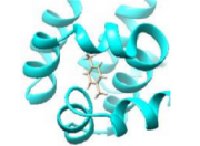
Predict a binding pose
Supply a protein and ligand, choose a binding region, then explore and download predicted poses. The live calculation uses AutoDock Vina through Webina on your computer. ETDock’s geometric approach is described in the research below.
From separate structures to a binding pose
A ligand’s chemical identity does not specify where it sits or how it is oriented in a protein. ETDock predicts distances between the binding partners, then uses those distances and constraints on the ligand’s internal geometry to reconstruct a pose.
The model combines atom-level and whole-molecule ligand features with a geometric protein representation. Its TAMformer module exchanges information through triangle, attention and message layers. Scalar features describe chemical properties; equivariant vectors preserve how spatial relationships change when a structure is rotated.
How closely do the predicted poses match?
The paper evaluates ETDock on a time split of PDBbind v2020. Ligand RMSD measures how closely predicted atom positions match the reference pose; lower values indicate a closer match.
- Median ligand RMSD
- 3.8 Å
- Poses within 2 Å
- 23.2%
- Poses within 5 Å
- 61.1%
Reported in Table 1 of the ETDock paper. These metrics describe pose accuracy in that evaluation; they do not measure binding affinity.
From a pose to an affinity estimate
A pose describes an arrangement of atoms. Affinity concerns the strength of association between the protein and ligand. The arrangement establishes the contacts that a structure-based affinity model can examine.
Study the binding contacts
AGIMA-Score investigates how graphs of protein–ligand contacts can inform affinity estimation from a complex.
Explore AGIMA-ScoreExplore a complex of your own
Already have a bound complex? The live GNINA tool scores the pose in your PDB structure and lets you explore its spatial occlusion response.
Score a bound complexPaper & resources
Yiqiang Yi, Yuting Huang, Xu Wan, Yatao Bian, Debby D. Wang, Peilin Zhao and Le Ou-Yang. ETDock: an equivariant transformer for protein-ligand docking. Frontiers of Computer Science 21(5), 2105903 (2027). Published online on 5 January 2026; assigned to the 15 May 2027 journal issue.
Yuri Kochnev, Erich Hellemann, Kevin C. Cassidy and Jacob D. Durrant. Webina: an open-source library and web app that runs AutoDock Vina entirely in the web browser. Bioinformatics 36(16), 4513–4515 (2020).
Oleg Trott and Arthur J. Olson. AutoDock Vina: Improving the speed and accuracy of docking with a new scoring function, efficient optimization, and multithreading. Journal of Computational Chemistry 31(2), 455–461 (2010).
Andrew T. McNutt, Yanjing Li, Rocco Meli, Rishal Aggarwal and David Ryan Koes. GNINA 1.3: the next increment in molecular docking with deep learning. Journal of Cheminformatics 17, 28 (2025).